Dental Cone Beam CT
Dental cone beam computed tomography (CT) is a special type of x-ray equipment used when regular dental or facial x-rays are not sufficient. Your doctor may use this technology to produce three dimensional (3-D) images of your teeth, soft tissues, nerve pathways and bone in a single scan.
This procedure requires little to no special preparation. Tell your doctor if there's a possibility you are pregnant. Wear loose, comfortable clothing and leave jewelry at home. You may be asked to wear a gown.
What are some common uses of the procedure?
Dental cone beam CT is commonly used for treatment planning of orthodontic issues. It is also useful for more complex cases that involve:
- surgical planning for impacted teeth.
- diagnosing temporomandibular joint disorder (TMJ).
- accurate placement of dental implants.
- evaluation of the jaw, sinuses, nerve canals and nasal cavity.
- detecting, measuring and treating jaw tumors.
- determining bone structure and tooth orientation.
- locating the origin of pain or pathology.
- cephalometric analysis.
- reconstructive surgery.
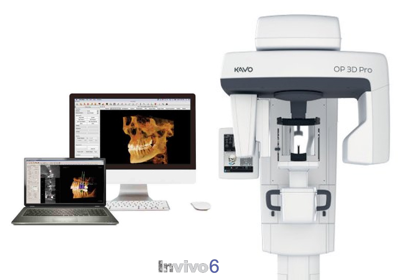
What does the equipment look like?
Cone beam CT scanners are square-shaped machines that include either an upright chair for sitting or a moveable table so patients can lie down during the examination. Scanners that include a chair have a rotating C-arm, an x-ray image intensifier that contains an x-ray source and detector. Cone beam CT machines with a table include a rotating gantry.
How does the procedure work?
During a cone beam CT examination, the C-arm or gantry rotates around the head in a complete 360-degree rotation while capturing multiple images from different angles that are reconstructed to create a single 3-D image.
The x-ray source and detector are mounted on opposite sides of the revolving C-arm or gantry and rotate in unison. In a single rotation, the detector can generate anywhere between 150 to 200 high resolution two-dimensional (2-D) images, which are then digitally combined to form a 3-D image that can provide your dentist or oral surgeon with valuable information about your oral and craniofacial health.
Who interprets the results and how do I get them?
Your dentist, oral surgeon or radiologist will analyze the images. They may discuss the results with you directly or communicate the results to your referring physician or dentist.
Reference
- https://www.radiologyinfo.org/en/info.cfm?pg=dentalconect
Reference
- https://www.kavo.com/en-us/imaging-solutions/kavo-op-3d-an-upgradeable-pan-panoramic-xray#overview